

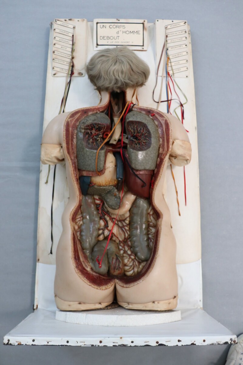
This educational anatomical model depicts a male torso in an upright position. An opening at the back provides a view of the internal organs of the respiratory, digestive and vascular systems. Made primarily from wax coloured throughout, it is enhanced with natural hair, adding to its lifelike appearance.
Originally designed as a teaching aid for medical students, the model made it possible to examine the internal organs and gain a better understanding of their anatomical relationships. With the rise of photography in the 19th century, such models also found their way into fairgrounds, helping to spread knowledge of the human body among the general public. This particular model was notably exhibited at the Brussels Midi Fair.
To facilitate the identification of the different anatomical structures, each organ is connected by a coloured thread to a numbered label. Today, this remarkable scientific artefact forms part of the permanent collection of the Museum of Medicine, where it continues to raise visitors’ awareness of human anatomy and how the body works.
However, the model is now in urgent need of restoration. It has suffered several significant fractures, particularly around the head and neck, which have become completely detached from the rest of the body. Furthermore, the artefact is made of a variety of materials (wax, natural hair, textiles and other elements), each showing specific forms of deterioration. Without conservation and restoration work, this precious testament to 19th-century scientific heritage risks suffering irreversible damage.
“This 200-year-old anatomical model continues to demonstrate its educational value to audiences of all kinds.”
— Marine Roosens, Director of the Museum of Medicine